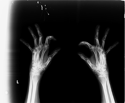
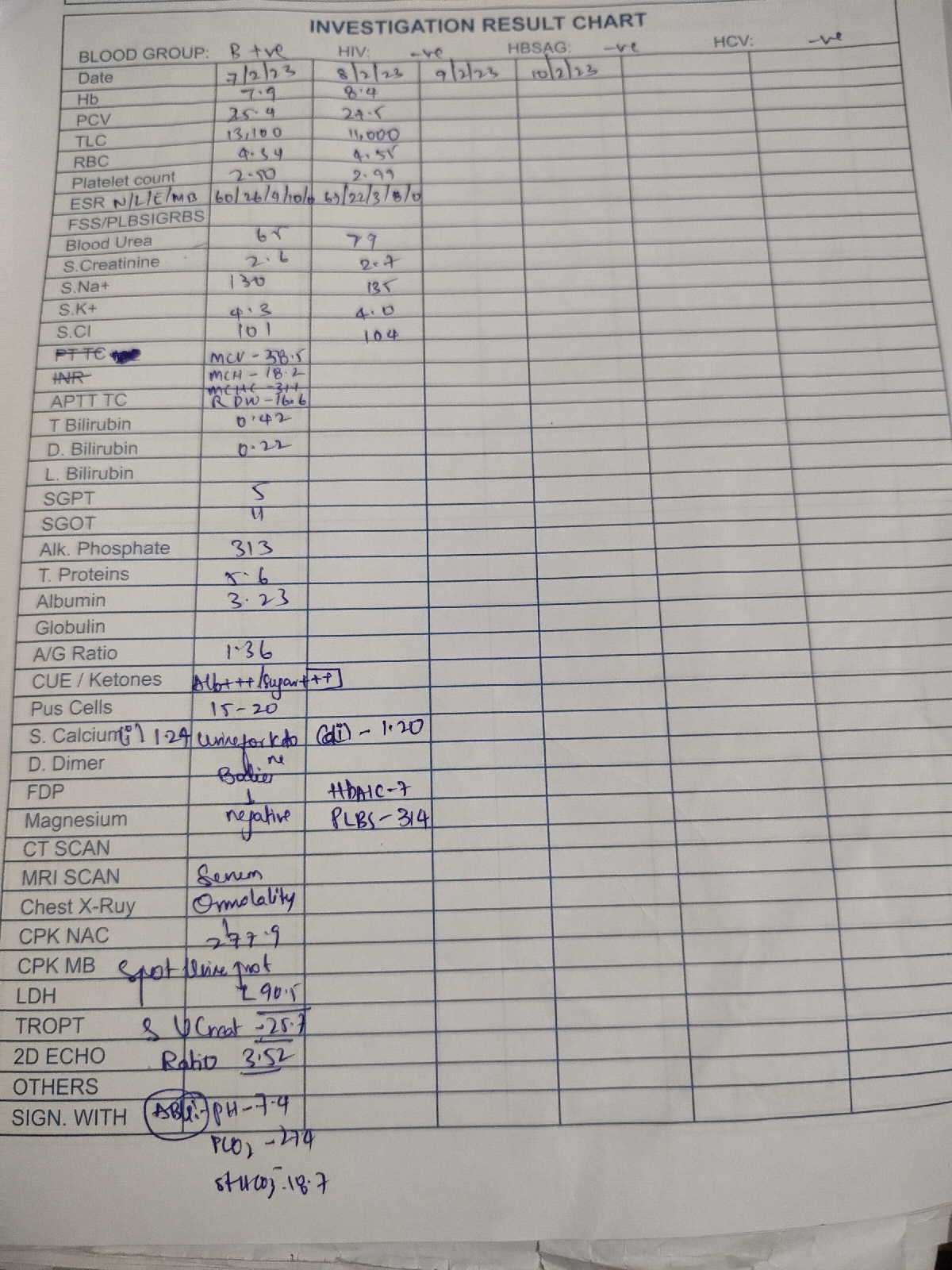
47 Year old female patient with fever and joint pains ( short case)
This is an E log book to discuss our patient's de-identified health data shared after taking his guardian's signed informed consent. Here we discuss our individual patient problems through series of inputs from available global online community of experts with an aim to solve those patient's clinical problems with collective current best evidence based inputs. This e-log book also reflects my patient centered online learning portfolio and your valuable comments in comment box are most welcomed
.
I have been given this case to solve in an attempt to understand the topic of "Patient clinical data analysis" to develop my competency i reading and comprehending clinical data including history, clinical findings, investigations and come up with a diagnosis and treatment plan.
H no : 1701006072
A 47 year old female tailor by occupation resident of nalgonda came to the OPD on 2_06_2022 with the chief complaints of
Fever since 3 months
Facial rash from 15 days
History of presenting illness:
Patient apparently asymptomatic 10 years back later she developed joint pains (in ankle and knee) it was associated with morning stiffness and limitation of joint movement . This get usually relieved after some activity .
For joint pains she went to local hospital where she tested RA positive
She was on diclofenac for 2 months and symptoms relieved
Last year she took COVID vaccination
Later she developed joint pains
After which she consulted orthopaedician and symptoms relieved by taking medication
3months back she had joint pains and fever which was Insidious in onset Intermittent on and off not associated with chills and rigor.
She went to the private hospital but the fever was recurrent associated with abdominal pain came here on 2/6/22
Patient also had facial rash over the face which increased on exposure to sun. It was a diffuse erythematous lesion and hyperpigmented papules were noted over the bilateral cheek sparing nasolabial folds and it developed from last 15 days
Past history
Patient had an history of gradual painless loss of vision since 2011and was certified as blind 2 years back
Not a known case of diabetes asthma Epilepsy thyroid tuberculosis and coronary artery disease.
No similar complaints in the family
Personal history
General examination
Pateint is consious coherent co operative well oriented to time place and person,moderately built and moderately nourished and is examined with informed consent.
Pallor: present
No icterus, cyanosis, clubbing,lymphadenopathy, edema.
VITALS
PULSE :86BPM
BP:120/80mm hg
RR:16cpm
SPO2:98%at room air
SYSTEMIC EXAMINATION
CVS
Inspection:SHAPE OF THE CHEST IS NORMAL
no visible neck veins
No rise in JVP
No visible pulsation scars.
Palpation
ALL inspectory findings are confirmed
Cardiac impulse felt at 5ty intercostal space 1cm medial to the mid clavicular line.
Percussion shows normal heart borders
Auscultation: s1 s2 heard no murmurs
CNS
Higher mental function normal
Cranial nerve examination normal
Normal motar and sensory system on examination
Respiratory examination
Inspection
Shape of chest is elliptical,
B/L symmetrical chest,
Trachea in central position,
Expansion of chest- normal on both sides
Palpation
All inspectory findings are confirmed,
No tenderness, No local rise of temperature,
Percussion
normal resonant note present bilaterally
auscultation: normal vesicular breath sounds heard
GIT
inspection- normal scaphoid abdomen with no pulsations and scars
palpation - inspectory findings are confirmed
no organomegaly, non tender and soft
percussion- normal resonant note present, liver border normal
auscultation-normal abdominal sounds heard, no bruit present
CBP
- Hemoglobin- 6 gm/dl
- PCV- 21 %
- TLC- 8200/ cumm
- RBC- 2.5 million/cumm
- Platelets- 1.32 lakhs/ml
CBP
- Hemoglobin- 6 gm/dl
- PCV- 21 %
- TLC- 8200/ cumm
- RBC- 2.5 million/cumm
- Platelets- 1.32 lakhs/ml
Chest x ray pA view
Ophthalmology report
Bilateral optic atrophy
Treatment given
PROVISIONAL DIAGNOSIS:
SECONDARY SJOGRENS SYNDROME
LEFT LOWER LIMB CELLULITIS WITH BILATERAL OPTIC ATROPHY.


